


Prescribing information (package insert) - Pixclara
1 INDICATIONS AND USAGE
PIXCLARA is indicated for use with positron emission tomography (PET) to differentiate recurrent or progressive glioma from treatment-related change, in conjunction with other diagnostic evaluations, in adults and pediatric patients 1 month of age and older.
2 DOSAGE AND ADMINISTRATION
2.1 Radiation Safety - Drug Handling
Handle PIXCLARA with appropriate safety measures to minimize radiation exposure [see Warnings and Precautions (5.2)]. Use waterproof gloves, effective radiation shielding, and other appropriate safety measures when preparing and handling PIXCLARA.
Radiopharmaceuticals should be used by or under the control of healthcare providers who are qualified by specific training and experience in the safe use and handling of radionuclides, and whose experience and training have been approved by the appropriate governmental agency authorized to license the use of radionuclides.
2.2 Recommended Dosage and Administration Instructions
Recommended Dosage in Adult Patients
The recommended amount of radioactivity for adults is 185 MBq to 259 MBq (5 mCi to 7 mCi) administered as an intravenous bolus injection.
Recommended Dosage in Pediatric Patients 1 Month of Age and Older
The recommended amount of radioactivity for pediatric patients 1 month of age and older is 3 MBq/kg actual body weight (0.081 mCi/kg) with a minimum and maximum activity of 14 MBq (0.38 mCi) and 259 MBq (7 mCi), respectively, administered as an intravenous bolus injection.
Administration
- Use aseptic technique and radiation shielding when withdrawing and administering PIXCLARA.
- Calculate the necessary volume to administer based on calibration time and required dose.
- Inspect PIXCLARA visually for particulate matter and discoloration before administration. Only use solutions that are clear, colorless or slightly yellow, and without visible particles.
- PIXCLARA may be diluted with 0.9% Sodium Chloride Injection.
- Assay the final dose in a dose calibrator immediately before administration to the patient.
- After injection of PIXCLARA, administer an intravenous flush of 0.9% Sodium Chloride Injection to ensure full delivery of the dose.
- Dispose of any unused drug in a safe manner in compliance with applicable regulations.
2.3 Patient Preparation
- Instruct patients to fast for at least 4 hours prior to administration of PIXCLARA.
- Encourage patients to drink water during the fasting period and after administration to promote adequate hydration, and to void, if possible, immediately prior to imaging and frequently after imaging [see Warnings and Precautions (5.2)].
2.4 Image Acquisition
- Position the patient with their head in a dedicated holder and their arms resting at their sides. Use tape, padding, or other flexible head restraints to minimize head movement.
- Begin PET image acquisition 20 minutes to 30 minutes after PIXCLARA administration. Acquire images for a single bed position centered on the brain for a duration of 20 minutes.
- A low dose computed tomography (CT) or magnetic resonance imaging (MRI) scan of the brain may be used for attenuation correction.
2.5 Image Display and Assessment
Image Display and Orientation
- Display images in all three anatomical planes: transverse (axial), sagittal, and coronal.
- Reorient images in all three planes to correct any head tilt.
- Fuse with a recent anatomic scan for better localization of tracer uptake.
- Perform all subsequent lesion selection and background measurements on transverse (axial) images.
Defining the Background SUV mean
- Identify the Primary Lesion: Locate the largest suspected lesion and identify the single slice that contains the voxel with the highest activity (maximal uptake).
Draw the Background Region of Interest (ROI):
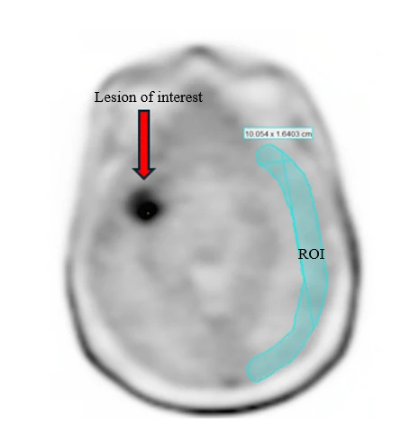
- On the identified slice, as well as the slices immediately above and below it, draw a crescent-shaped (banana-shaped) ROI in the contralateral (unaffected) hemisphere (see Figure 1).
- The ROI should be 1.5 cm to 2 cm in width and positioned at the periphery of the brain to contain primarily cortical gray matter.
- Refine the Background ROI: Adjust the ROI to ensure it only contains normal gray and white matter. Exclude any abnormal brain tissue such as prior infarct and regions suspected of prior surgery. Also exclude normal structures such as the ventricular system, venous sinuses, and skull.
- Measure the Background Intensity: Calculate the mean Standardized Uptake Value (SUVmean) in the background ROI (Background SUVmean).
Figure 1: Selection of Background ROI. Place a 1.5 cm to 2 cm wide crescent-shaped ROI on the contralateral side to the lesion of interest encompassing predominantly gray matter, avoiding inclusion of skull and blood pool.
Setting the Color Scale for Visual Assessment
- Select an Appropriate Color Scale: Utilize a color scale with a rapid transition between two distinct colors. The Sokoloff scale, which transitions from blue to green at 50% of maximum contrast, is a suitable example.
- Set the Color Transition Point: Adjust the color scale so that the color transition occurs at 1.6 times the Background SUVmean.
Set the Upper Contrast Value (UCV): Set the upper limit of the visual intensity scale using the following formula:
- UCV = (Background SUVmean × 1.6) × (100% / % level of color transition)
- Example: For the Sokoloff scale, the transition is at 50%. The formula would be: UCV = (Background SUVmean × 1.6) × (100% / 50%), or (Background SUVmean × 1.6) × 2.
- Final Visual Setup: Once set, normal brain activity will appear in the lower range of the color scale while regions suspicious for disease will appear in the upper range, allowing for visual assessment.
Calculating the Target-to-Background Ratio (TBR) for Lesions
This step provides semi-quantitative metrics for areas suspected of disease recurrence or progression. Two distinct measurements are required.
Whole Lesion Analysis (for TBRmean)
- 1. Create a Threshold Volume of Interest (VOI): Contour a VOI that encompasses all increased uptake within the lesion. Apply a threshold to include only voxels with an SUV greater than background SUVmean × 1.6.
- 2. Refine the VOI: Manually edit the VOI to exclude any area of physiologic uptake (e.g., skull, venous sinus blood pool).
- 3.
Calculate TBRmean: Use the SUVmean from this refined VOI to calculate the mean Target-to-Background Ratio (TBRmean) with the following formula:
- TBRmean = SUVmean of Threshold VOI / Background SUVmean
Hottest Spot Analysis (for TBRmax)
- 1. Locate the Hottest Voxel: Within the Threshold VOI created above, navigate to the single voxel with the maximum SUV (SUVmax).
- 2. Place a Circular ROI: Center a circular ROI with 1.6 cm diameter on this SUVmax voxel.
- 3. Adjust the ROI: If necessary, move the circular ROI to maximize the amount of high-uptake voxels it contains.
- 4.
Calculate TBRmax: Use the SUVmean from this 1.6 cm circular ROI to calculate the maximum Target-to-Background Ratio (TBRmax) with the following formula:
- TBRmax = SUVmean of 1.6 cm circle ROI / Background SUVmean
2.6 Image Interpretation
General Principles of Interpretation
Assess each suspicious region on a PIXCLARA PET scan to differentiate recurrence or progression from treatment-related change.
- Positive PIXCLARA uptake: A region with positive uptake is concerning for progression or recurrence.
- Negative PIXCLARA uptake: A region with negative uptake is more likely to represent a treatment-related change (e.g., pseudoprogression, radiation necrosis).
- Images should be interpreted by first performing a visual assessment, then evaluating semi-quantitative PIXCLARA PET tumor-to-background ratio thresholds (TBRmean and TBRmax), finally integrating both visual and semi-quantitative findings to determine the overall interpretation [see Dosage and Administration (2.5)]. Do not rely solely on semi-quantitative values for interpretation.
Positive PIXCLARA Images
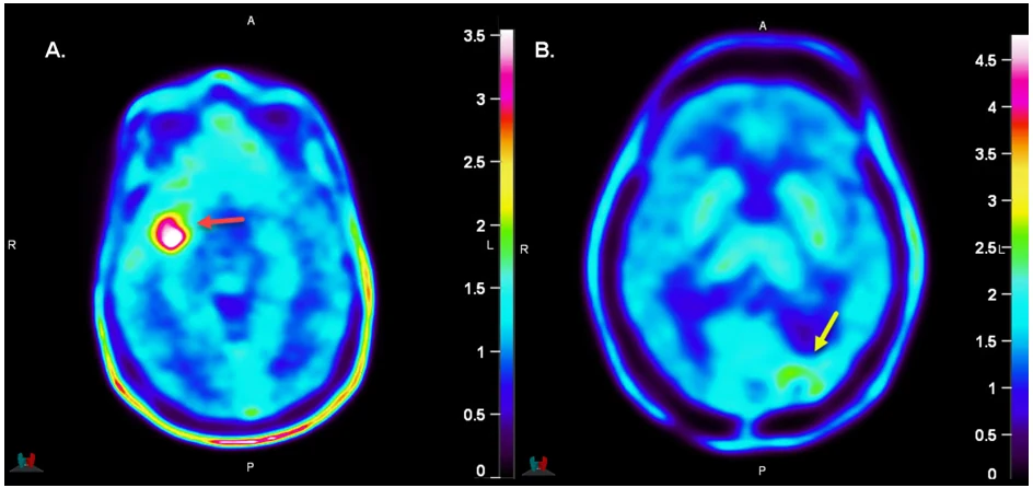
A lesion is considered positive based on the following criteria. (See Figure 2A for an example of a positive lesion.)
- Visual Assessment: A visually positive lesion demonstrates a cluster of contiguous voxels with uptake significantly above the surrounding background brain tissue. When using the Sokoloff color scale with recommended settings, this increased uptake appears in the yellow, orange, or red range [see Dosage and Administration (2.5)]. Evaluate other visual features such as the focality and morphology of the uptake in addition to uptake intensity.
- Semi-Quantitative Assessment: Values of TBRmean ≥ 2 and TBRmax ≥ 2.3 are suggestive of positive PIXCLARA uptake.
Negative PIXCLARA Images
A lesion is considered negative based on the following criteria. (See Figure 2B for an example of a negative lesion.)
- Visual Assessment: A visually negative lesion shows no voxels with uptake above the background. When using the Sokoloff color scale with recommended settings, voxels will be in the blue and light blue color range. A lesion is likely negative if a VOI cannot be contoured with uptake greater than 1.6 times the SUVmean of the background.
- Semi-Quantitative Assessment: TBRmean <2 and TBRmax <2.3 are suggestive of negative PIXCLARA uptake.
Factors Affecting Interpretation and Potential for Inaccurate Results
Potential for False Positive Results:
- Inflammation, infection, and other non-neoplastic inflammatory processes can cause increased uptake of floretyrosine F 18, potentially leading to false positive results.
- Since floretyrosine F 18 remains in the blood pool at the time of imaging, increased signal may be observed in large vessels (e.g., sagittal, transverse, and cavernous sinus) and vascular malformations (e.g., arterio-venous malformations, angiomas, cavernomas, and developmental venous anomalies), which can also cause false positive results.
Potential for False Negative Results:
- Tumor heterogeneity can present a challenge, as different parts of a tumor may have varying levels of uptake. Areas with lower uptake could be misinterpreted as non-tumorous, leading to a false negative result.
- Small lesions may be subject to partial volume effects, which can cause an underestimation of the true uptake and lead to a negative result by semi-quantitative assessment even if the lesion is malignant.
- Readers should always consider other factors that may impact semi-quantitative assessments, such as variability in background uptake due to conditions like brain atrophy.
Figure 2: Examples of Positive and Negative PIXCLARA Images.
A. Positive Image: A patient with a treated glioblastoma in the right temporal lobe, showing positive visual uptake, TBRmean = 2.2, and TBRmax = 2.9. Axial image using the Sokoloff color scale, with upper limit set to SUV 3.5 (transition from blue to green occurs at SUV 1.8). B. Negative Image: A patient with a treated grade III diffuse astrocytoma in the left occipital lobe showing negative visual uptake, TBRmean = 1.7, and TBRmax = 1.5. Axial image using the Sokoloff color scale, with upper limit set to SUV 4.6 (transition from blue to green occurs at SUV 2.3).
2.7 Radiation Dosimetry
Estimated radiation absorbed doses per injected activity for organs and tissues of adult patients following an intravenous bolus of PIXCLARA are shown in Table 1.
The effective dose resulting from the administration of 259 MBq of PIXCLARA in an adult is about 4.1 mSv. For an administered activity of 259 MBq, the urinary bladder wall had the highest absorbed dose of 22 mGy.
These radiation doses are for PIXCLARA alone. If CT is used for attenuation correction, the radiation dose will increase by an amount that varies by technique.
Table 1: Estimated Radiation Absorbed Dose per Injected Activity in Selected Organs and Tissues of Adults and Pediatric Patients after Intravenous Administration of PIXCLARA
| Organ/Tissue | Absorbed Dose per Unit Activity Administered (mGy/MBq) | ||||
|---|---|---|---|---|---|
| Adults | 15 years | 10 years | 5 years | 1 year | |
| Adrenals | 0.014 | 0.017 | 0.026 | 0.042 | 0.077 |
| Bone surfaces | 0.013 | 0.016 | 0.024 | 0.039 | 0.074 |
| Brain | 0.01 | 0.013 | 0.021 | 0.034 | 0.064 |
| Breasts | 0.0095 | 0.012 | 0.018 | 0.03 | 0.057 |
| Colon | 0.011 | 0.013 | 0.021 | 0.032 | 0.054 |
| Esophagus | 0.012 | 0.015 | 0.023 | 0.036 | 0.069 |
| Gallbladder Wall | 0.014 | 0.017 | 0.026 | 0.038 | 0.068 |
| Heart Wall | 0.013 | 0.016 | 0.026 | 0.039 | 0.072 |
| Kidneys | 0.027 | 0.033 | 0.046 | 0.069 | 0.12 |
| Liver | 0.017 | 0.022 | 0.032 | 0.048 | 0.088 |
| Lower Large Intestine Wall | 0.012 | 0.014 | 0.022 | 0.033 | 0.054 |
| Lungs | 0.014 | 0.02 | 0.028 | 0.042 | 0.081 |
| Muscles | 0.012 | 0.014 | 0.023 | 0.036 | 0.067 |
| Ovaries | 0.015 | 0.018 | 0.028 | 0.043 | 0.077 |
| Pancreas | 0.014 | 0.018 | 0.027 | 0.043 | 0.078 |
| Red Bone Marrow | 0.013 | 0.016 | 0.024 | 0.038 | 0.072 |
| Skin | 0.009 | 0.011 | 0.018 | 0.029 | 0.055 |
| Small Intestine Wall | 0.0076 | 0.0094 | 0.014 | 0.02 | 0.032 |
| Spleen | 0.013 | 0.016 | 0.024 | 0.04 | 0.073 |
| Stomach Wall | 0.013 | 0.016 | 0.024 | 0.038 | 0.069 |
| Testes | 0.012 | 0.016 | 0.025 | 0.038 | 0.07 |
| Thymus | 0.012 | 0.015 | 0.023 | 0.036 | 0.069 |
| Thyroid | 0.012 | 0.015 | 0.024 | 0.039 | 0.073 |
| Upper Large Intestine Wall | 0.01 | 0.013 | 0.02 | 0.031 | 0.054 |
| Urinary bladder wall | 0.085 | 0.11 | 0.16 | 0.22 | 0.3 |
| Uterus | 0.017 | 0.021 | 0.034 | 0.051 | 0.086 |
| Remaining Organs | 0.012 | 0.014 | 0.022 | 0.035 | 0.066 |
| Effective Dose (mSv/MBq) | 0.016 | 0.021 | 0.031 | 0.047 | 0.082 |
3 DOSAGE FORMS AND STRENGTHS
Injection: 185 MBq/mL to 11,100 MBq/mL (5 mCi/mL to 300 mCi/mL) of floretyrosine F 18 in approximately 15 mL at the end of synthesis (EOS) as a clear, colorless to slightly yellow solution in a multiple-dose vial.
4 CONTRAINDICATIONS
None
5 WARNINGS AND PRECAUTIONS
5.1 Risk for Misinterpretation
Image misinterpretation may occur with PIXCLARA PET. A negative image does not rule out the presence of recurrent or progressive glioma and a positive image does not confirm the presence of recurrent or progressive glioma. Equivocal findings may occur with PIXCLARA PET, including low-level or atypical uptake patterns, which may result in false positive or false negative interpretations [see Dosage and Administration (2.6)].
Interpret PIXCLARA PET findings with caution and correlate results with other available diagnostic evaluations, such as histopathology, cross-sectional imaging, and clinical history, to support appropriate clinical decision-making.
5.2 Radiation Risks
PIXCLARA contributes to a patient's overall long-term cumulative radiation exposure. Long-term cumulative radiation exposure is associated with an increased risk for cancer. Ensure safe drug handling to protect patients and health care providers from unintentional radiation exposure. Advise patients to hydrate before and after administration and to void frequently after administration [see Dosage and Administration (2.1, 2.3)].
6 ADVERSE REACTIONS
6.1 Clinical Trials Experience
Because clinical trials are conducted under widely varying conditions, adverse reaction rates observed in the clinical trials of a drug cannot be directly compared to rates in the clinical trials of another drug and may not reflect the rates observed in practice.
The safety of PIXCLARA was evaluated in 382 patients with gliomas. Among these 382 patients, 371 adult patients received at least one intravenous dose of PIXCLARA at a mean activity of 204 ± 18 MBq (5.5 ± 0.47 mCi). The remaining 11 pediatric patients received at least one intravenous dose of PIXCLARA at a mean activity of 155 ± 53.5 MBq (4.2 ± 1.4 mCi).
The mean age of the patients was 57 years (range: 5 years to 86 years). Sex was 59% male, 37% female, and 3% unreported. Distribution by race was 26% White, 6% Asian, <1% Black or African American, and 67% other or unreported. Distribution by ethnicity was 3% Hispanic/Latino, 96% non-Hispanic/Latino, and <1% unknown or unreported.
Adverse reactions that occurred in ≥0.5% of patients receiving PIXCLARA were headache (0.5%).
Adverse reactions that occurred in <0.5% of patients were nausea, injection site reaction, fatigue, and malaise.
Adverse Reactions in Pediatric Patients
Overall, the safety profile observed in pediatric patients from the clinical study was consistent with the safety profile in adult patients [see Use in Specific Populations (8.4) and Clinical Studies (14)].
8 USE IN SPECIFIC POPULATIONS
8.1 Pregnancy
Risk Summary
There are no data on use of floretyrosine F 18 in pregnant women to evaluate for a drug-associated risk of major birth defects, miscarriage, or other adverse maternal or fetal outcomes.
All radiopharmaceuticals, including PIXCLARA, have the potential to cause fetal harm depending on the fetal stage of development and the magnitude of radiation dose. If considering PIXCLARA administration to a pregnant woman, inform the patient about the potential for adverse pregnancy outcomes based on the radiation dose from PIXCLARA and the gestational timing of exposure.
The background risk of major birth defects and miscarriage for the indicated populations is unknown. All pregnancies have a background risk of birth defects, loss, or other adverse outcomes. In the U.S. general population, the estimated background risk of major birth defects and miscarriage in clinically recognized pregnancies is 2% to 4% and 15% to 20%, respectively.
8.2 Lactation
Risk Summary
There are no data on the presence of floretyrosine F 18 in human or animal milk, the effects on the breastfed infant, or effects on milk production. Exposure of floretyrosine F 18 to a breastfed infant can be minimized by advising a lactating woman to temporarily discontinue breastfeeding and to pump and discard breast milk for a minimum of 4 hours after administration of PIXCLARA. The developmental and health benefits of breastfeeding should be considered along with the mother's clinical need for PIXCLARA and any potential adverse effects on the breastfed child from PIXCLARA or from the underlying maternal condition.
8.4 Pediatric Use
The safety and effectiveness of PIXCLARA have been established in pediatric patients 1 month of age and older. Use of PIXCLARA in this age group is supported by evidence from adequate and well-controlled studies of PIXCLARA in adults and pediatric patients 5 to 16 years of age [see Adverse Reactions (6) and Clinical Studies (14)] with additional data from published literature supporting the safety and effectiveness of weight-based dosing of floretyrosine F 18 in glioma imaging.
The safety and effectiveness of PIXCLARA have not been established in pediatric patients younger than 1 month of age.
8.5 Geriatric Use
Of the total number of subjects in clinical studies of PIXCLARA, 180 (28.3%) were 65 years of age and over, while 51 (8.0%) were 75 years of age and over. No overall differences in safety or effectiveness were observed between these subjects and younger subjects, and other reported clinical experience has not identified differences between elderly and younger patients.
10 OVERDOSAGE
In the event of an overdose of PIXCLARA, reduce the radiation absorbed dose to the patient when possible by increasing the elimination of the drug from the body using hydration and frequent bladder voiding. A diuretic might also be considered. If possible, an estimate of the radiation effective dose given to the patient should be made.
11 DESCRIPTION
11.1 Drug Characteristics
PIXCLARA (floretyrosine F 18) injection is a radioactive diagnostic drug for intravenous use.
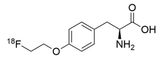
Floretyrosine F 18 is a radiolabeled derivative of the amino acid, tyrosine. Floretyrosine F 18 is chemically (2S)-2-amino-3-[4-(2-(18F)fluoroethoxy)phenyl]propanoic acid and has the molecular formula C11H14 18FNO3, molecular weight of 226.2 g/mol, and chemical structure as shown in Figure 3.
| Figure 3: Chemical Structure of Floretyrosine F 18 |
|---|
 |
PIXCLARA is a sterile, clear, colorless to slightly yellow solution with a pH between 4.5 and 6.5. Each mL contains no more than 8.33 mcg floretyrosine, 185 MBq to 11,100 MBq (5 mCi to 300 mCi) floretyrosine F 18 at the end of synthesis (EOS), and the inactive ingredient 0.5 mg sodium ascorbate in 0.9% sodium chloride injection.
11.2 Nuclear Physical Characteristics
Fluorine-18 (18F) decays by positron emission to stable oxygen-18 with a half-life of 109.8 minutes. The principal photons useful for diagnostic imaging are the coincident pair of 511 keV gamma photons, resulting from the interaction of an emitted positron with an electron (see Table 2).
Table 2: Principal Radiation Produced from Decay of Fluorine-18
| Radiation/Emission | % Per Disintegration | Mean Energy (keV) |
|---|---|---|
| Positron | 96.7 | 249.8 |
| Gamma | 193.5 | 511 |
The point source air kerma coefficient for 18F is 3.75 × 10⁻¹⁷ Gy m2/(Bq s) at 1 cm. The first half-value layer of lead (Pb) for the 18F gamma rays is approximately 6 mm. The relative reduction of radiation emitted by 18F that results from various thicknesses of lead shielding is shown in Table 3. The use of 8 cm of Pb will decrease the radiation transmission (i.e., exposure) by a factor of about 10,000.
Table 3: Radiation Attenuation of 511 keV Gamma Rays by Lead Shielding
| Shield Thickness cm of Lead (Pb) | Coefficient of Attenuation |
|---|---|
| 0.6 | 0.5 |
| 2 | 0.1 |
| 4 | 0.01 |
| 6 | 0.001 |
| 8 | 0.0001 |
For use in correcting for physical decay of 18F, the fractions remaining at selected intervals after calibration are shown in Table 4.
Table 4: Physical Decay Chart for Fluorine-18
| Minutes | Fraction Remaining |
|---|---|
| 0 | 1 |
| 15 | 0.909 |
| 30 | 0.826 |
| 60 | 0.683 |
| 110 | 0.5 |
| 220 | 0.25 |
12 CLINICAL PHARMACOLOGY
12.1 Mechanism of Action
Floretyrosine F 18 is a radiolabeled derivative of tyrosine and a substrate for the Large Neutral Amino Acid Transporter 1 and 2 (LAT). LAT is a sodium-independent exchanger protein that transports large neutral amino acids (e.g., tyrosine, phenylalanine) across cell membranes, and it is highly expressed in the blood-brain barrier and glioma tumor tissue. Consequently, floretyrosine F 18 accumulates in cells that express LAT on their surface. Fluorine-18 is a β+ emitting radionuclide that enables positron emission tomography.
12.2 Pharmacodynamics
The relationship between floretyrosine F 18 plasma concentrations and image interpretation has not been characterized.
12.3 Pharmacokinetics
Distribution
Following intravenous administration, floretyrosine F 18 distributes (% of injected activity) to skeletal muscles (39.2%), liver (3.1%), lungs (2.3%), bone marrow (2.2%), and brain (1.9%).
Elimination
Metabolism
The majority of floretyrosine F 18 remains intact in plasma. At 60 minutes and 120 minutes post-injection, 91 ± 5% and 87% ± 13% of the total radioactivity in the plasma was intact floretyrosine F 18, respectively.
Excretion
Elimination is by urinary excretion. Approximately 22% of administered activity was excreted by 5 hours.
Drug Interaction Studies
In Vitro
- Effect of Transporters on Floretyrosine: Floretyrosine is not a substrate for P-glycoprotein (P-gp), breast cancer resistance protein (BCRP), or bile salt export pump (BSEP).
- Effect of Floretyrosine on CYP Enzymes: Floretyrosine does not inhibit or induce CYP1A2, CYP2B6, or CYP3A4.
- Effect of Floretyrosine on Transporter Systems: Floretyrosine does not inhibit P-gp, BCRP, or BSEP.
13 NONCLINICAL TOXICOLOGY
13.1 Carcinogenesis, Mutagenesis, Impairment of Fertility
Animal studies to assess the carcinogenicity or mutagenic potential of floretyrosine have not been conducted. However, floretyrosine has the potential to be mutagenic when radiolabeled with fluorine-18. No animal studies with floretyrosine have been performed to evaluate the potential for impairment of fertility in males or females.
14 CLINICAL STUDIES
The effectiveness of PIXCLARA in differentiating recurrent or progressive glioma from treatment-related change was evaluated in two clinical studies: a prospective, single center, open-label study (Study 1; NCT04044937), and a blinded central read study (Study 2) analyzing retrospectively collected images from multicenter, real-world clinical practice.
Study 1
Patients enrolled in Study 1 presented with intracranial neoplasm after primary treatment with radiation therapy, surgery, or both, and had suspected disease progression or recurrence based on MRI. The image analysis included a total of 127 patients (116 adults, 7 adolescents aged 12 years to 16 years, and 4 children aged 5 years to 11 years) with suspected recurrent or progressive glioma of any grade. Adult patients received 217 ± 23 MBq (5.9 ± 0.63 mCi) of PIXCLARA and pediatric patients received 155 ± 54 MBq (4.2 ± 1.4 mCi) followed by PET/MR imaging of the brain.
Three independent nuclear medicine experts performed blinded reads of the PIXCLARA PET images. Readers were blinded to all other clinical and imaging data except non-contrast T1 MR images used for localization of PET findings. Readers were instructed to identify up to five lesions and assign each as tumor (positive) or treatment-related change (negative) [see Dosage and Administration (2.5, 2.6)].
A multidisciplinary panel of experts determined the composite reference standard results. This panel reviewed any MR images obtained before, with, and up to 6 months after PIXCLARA PET, histopathology reports, additional conventional imaging reports, clinical reports, and surgical reports and assessed each lesion as tumor (positive) or treatment-related change (negative) based on totality of the available evidence. The panel did not have access to the blinded reader assessments or the PIXCLARA PET images.
The mean age of the patients in the efficacy analysis was 48 years (range 5 years to 85 years), and 62% were male. Of the study population, 73% were White, 14% Asian, <1% Black or African American, 12% of other or unknown race, and 8% Hispanic or Latino. Glioma grade was 1 (<1%), 2 (11%), 3 (21%), or 4 (67%). Isocitrate dehydrogenase was wild type in 60%, mutant in 31%, and not available in 9%. Bevacizumab was administered to 35% of patients within 6 months of PIXCLARA imaging. According to reference standard, 52% of patients had at least one lesion considered to be recurrent or progressive glioma.
PIXCLARA PET blinded reads were compared to the reference standard for detection of recurrent or progressive glioma at the lesion-level, and patient-level results, where at least one true positive lesion defined a true positive patient, are shown in Table 5.
Table 5: Lesion-Based, Patient-Level Performance of PIXCLARA PET for Detection of Recurrent or Progressive Glioma in Study 1 (n=127)For a given reader, the patient-level result is determined from the lesion-level result(s) using a composite reference standard.
| Endpoint | Reader 1 | Reader 2 | Reader 3 |
|---|---|---|---|
| Abbreviations: CI = confidence interval, n = number of patients, PPA = positive percent agreement, NPA = negative percent agreement | |||
| True Positive, n | 48 | 55 | 46 |
| True Negative, n | 36 | 29 | 39 |
| False Positive, n | 25 | 32 | 22 |
| False Negative, n | 18 | 11 | 20 |
PPAPPA represents the percentage of patients with a positive PIXCLARA PET result among those who were reference standard positive., % (95% CI) | 73 (61, 82) | 83 (73, 90) | 70 (58, 79) |
NPANPA represents the percentage of patients with a negative PIXCLARA PET result among those who were reference standard negative., % (95% CI) | 59 (47, 71) | 48 (36, 60) | 64 (51, 75) |
Among the 11 pediatric patients included in the study, positive percent agreement (PPA) was 83% (95% confidence interval (CI): 44%, 97%) and negative percent agreement (NPA) ranged from 40% (95% CI: 12%, 77%) to 60% (95% CI: 23%, 88%) among the three readers.
Study 2
Patients included in Study 2 had known or suspected glioma of any grade treated with surgery, radiation, or chemotherapy, and had suspected disease recurrence or progression based on MRI. The image analysis included a total of 254 patients (253 adults and 1 adolescent aged 16 years). Adult patients received 201 ± 22 MBq (5.4 ± 0.58 mCi) of PIXCLARA and the pediatric patient received 181 MBq (4.9 mCi) followed by PET/CT or PET/MR imaging of the brain.
Three independent nuclear medicine experts performed blinded reads of the PIXCLARA PET images. Readers were blinded to all other clinical and imaging data except non-contrast T1 MR images used for localization of PET findings. Readers were instructed to identify up to five lesions and assign each as tumor (positive) or treatment-related change (negative) [see Dosage and Administration (2.5, 2.6)].
A multidisciplinary panel of experts determined the composite reference standard results. This panel reviewed any MR images obtained before, with, and after PIXCLARA, histopathology reports, clinical reports, and surgical reports and assessed each lesion as tumor (positive) or treatment-related change (negative) based on totality of the available evidence. The panel did not have access to the blinded reader assessments or the PIXCLARA PET images.
The mean age of the patients in the efficacy analysis was 51 years (range 16 years to 83 years), and 58% were male. Of the study population, 20% were White, 2% Asian, and 78% of other or unknown race. Glioma grade was 1 (<1%), 2 (17%), 3 (15%), or 4 (67%). Isocitrate dehydrogenase was wild type in 4%, mutant in 9%, and other or not available in 87%. Bevacizumab was administered to 4% of patients within 6 months of PIXCLARA imaging. According to the reference standard, 50% of patients had at least one lesion considered to be recurrent or progressive glioma.
PIXCLARA PET blinded reads were compared to the reference standard for detection of recurrent or progressive glioma at the lesion-level, and patient-level results, where at least one true positive lesion defined a true positive patient, are shown in Table 6.
Table 6: Lesion-Based, Patient-Level Performance of PIXCLARA PET for Detection of Recurrent or Progressive Glioma in Study 2 (n=254)For a given reader, the patient-level result is determined from the lesion-level result(s) using a composite reference standard. These results are consistent with those of a tipping point analysis using p=0, i.e., all missing PET are imputed as negative. The numbers of reference-positive and reference-negative are consistent across the readers.
| Endpoint | Reader 1 | Reader 2 | Reader 3 |
|---|---|---|---|
| Abbreviations: CI = confidence interval, n = number of patients, PPA = positive percent agreement, NPA = negative percent agreement | |||
| True Positive, n | 84 | 92 | 85 |
| True Negative, n | 77 | 67 | 72 |
| False Positive, n | 50 | 60 | 55 |
| False Negative, n | 43 | 35 | 42 |
PPAPPA represents the percentage of patients with a positive PIXCLARA PET result among those who were reference standard positive., % (95% CI) | 66 (58, 74) | 72 (64, 79) | 67 (58, 75) |
NPANPA represents the percentage of patients with a negative PIXCLARA PET result among those who were reference standard negative., % (95% CI) | 61 (52, 69) | 53 (44, 61) | 57 (48, 65) |
16 HOW SUPPLIED/STORAGE AND HANDLING
How Supplied
PIXCLARA (floretyrosine F 18) injection is supplied at a concentration of 185 MBq/mL to 11,100 MBq/mL (5 mCi/mL to 300 mCi/mL) floretyrosine F 18 in approximately 15 mL at the end of synthesis as a clear, colorless to slightly yellow solution in a shielded multiple-dose glass vial (NDC# 74725-338-30).
Storage and Handling
Store PIXCLARA at 15°C to 25°C (59°F to 77°F) upright in the original container with radiation shielding.
Use PIXCLARA within 14 hours from the time of the end of synthesis. The expiration date and time are provided on the shield container label.
Dispose of any unused drug in accordance with applicable regulations.
This preparation is for use by persons under license by the Nuclear Regulatory Commission or the relevant regulatory authority of an Agreement State.
17 PATIENT COUNSELING INFORMATION
Radiation Risks
Advise patients of the radiation risks with PIXCLARA. Instruct patients to drink a sufficient amount of water to ensure adequate hydration before their PET study and urge them to drink and urinate frequently following the administration of PIXCLARA to reduce radiation exposure [see Dosage and Administration (2.3) and Warnings and Precautions (5.2)].
Fasting
Instruct patients to not eat for at least 4 hours prior to injection of PIXCLARA [see Dosage and Administration (2.3)].
Pregnancy
Inform pregnant women of the risk of fetal exposure to radiation doses if they undergo a radionuclide procedure with PIXCLARA [see Use in Specific Populations (8.1)].
Lactation
Advise a lactating woman to temporarily discontinue breastfeeding and to pump and discard breast milk for a minimum of 4 hours after PIXCLARA administration to minimize radiation exposure to the breastfed infant [see Use in Specific Populations (8.2)].
Manufactured for:
Telix Pharmaceuticals US, Inc.
11700 Exit 5 Pkwy, Suite 200
Fishers, IN 46037
USA
For optional information about PIXCLARA, contact Telix Pharmaceuticals (US) Inc. at 1-844-455-8638.
PIXCLARA is a registered trademark of Telix Pharmaceuticals Limited
Copyright © 2026 Telix Pharmaceuticals (US) Inc.
All rights reserved.